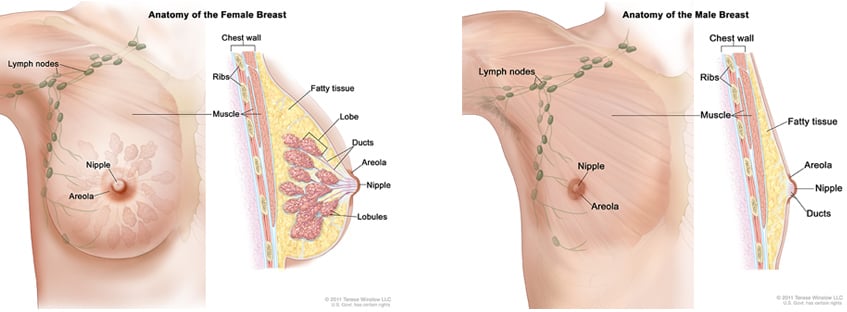
The Role of Lymph Nodes in Breast Cancer
One of the first places breast cancer can spread to and start growing in is the nearby lymph nodes. The lymphatic (lymph) system is part of the body’s immune system, which protects against infection and disease. This system is comprised of three elements, the lymph - a clear fluid that circulates through the lymphatic system, lymphatic vessels, and lymph nodes. The primary function of the lymph system is to circulate the lymph, which contains infection-fighting white blood cells, throughout the body to flush out toxins, waste, and other unwanted materials.
When breast cancer cells are multiplying, they can enter the lymphatic vessels located in a woman’s breast tissue. The lymph fluid then carries these cancerous cells throughout the body. The closest lymph nodes, usually in the underarm area, are often the first place that breast cancer will start to grow outside of the breast.
When an oncologist performs tests and determines that there are breast cancer cells in the lymph nodes, this is called lymph node involvement. Learn more about how breast cancer is detected and diagnosed.

Determining Lymph Node Involvement
To determine if lymph nodes are involved, your breast cancer specialist will remove one or several underarm lymph nodes so they can be examined under a microscope.
Lymph nodes can be checked in two different ways. The most common and least-invasive method is called sentinel lymph node biopsy. The other is called axillary lymph node dissection.
In most cases, lymph node surgery is done as part of the main surgery to remove the breast cancer. However, there are times it may be done as a separate operation.
Sentinel Lymph Node Biopsy (SLNB)
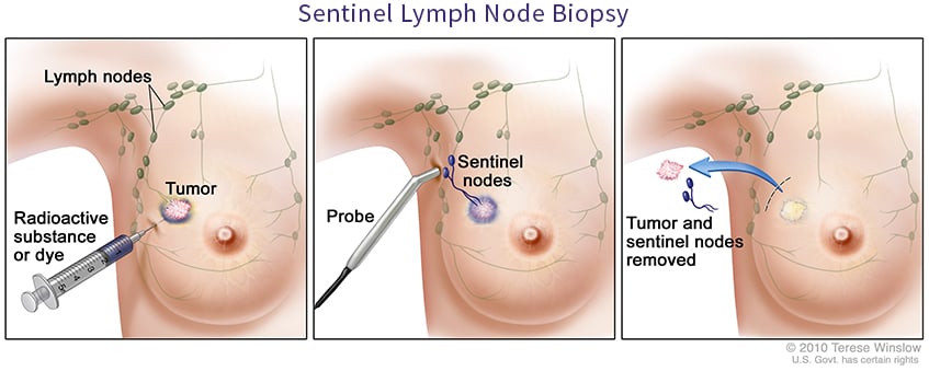
A sentinel lymph node is defined as the first lymph node to which cancer cells are most likely to spread from a primary tumor. Sometimes, there can be more than one sentinel lymph node.
During surgery to remove early-stage breast cancer, the sentinel node is identified and then removed so it can be sent to a pathologist (a physician who studies the causes and effects of diseases). The pathologist will determine if the biopsied lymph node is infected with cancer cells. The procedure to remove the sentinel lymph node for examination is called a sentinel lymph node biopsy (SLNB).

To identify the sentinel node:
- The surgeon injects either a radioactive substance, a blue dye, or both near the tumor.
- Depending on the substance that was injected, the surgeon then uses a device that detects radioactivity to find the sentinel node or looks for lymph nodes that are stained with the blue dye.
- Once the sentinel lymph node is located, the surgeon makes a small incision (about 1/2 inch) in the overlying skin and removes the node.
If the findings show no cancer in the sentinel nodes (lymph node-negative), then it is unlikely that other lymph nodes have cancer so surgery to remove more lymph nodes is not necessary. If cancer is found in the sentinel nodes (lymph node-positive), however, more lymph nodes may be removed with a procedure called axillary dissection.
Axillary Lymph Node Dissection (ALND)
The axillary lymph nodes run from the breast tissue into the armpit. This area under the arm is called the axilla.
During an axillary lymph node dissection, anywhere from 10 to 40 lymph nodes are removed and examined. These nodes are typically removed during your lumpectomy or mastectomy.
Lymph Node Status and Breast Cancer Treatment
The biopsy results (called a pathology report) will show how many lymph nodes were removed and how many were “involved” (tested positive for cancer). This is referred to as lymph node status. The results of the pathology exam help physicians determine the stage of breast cancer to recommend a treatment plan.
If the breast cancer has not spread to nearby lymph nodes, the status is referred to as node-negative. If the report indicates that cancer is present in the lymph nodes, the status is referred to as node-positive. Positive results also mean that the cancer may have already or could possibly spread to other organs, such as the bones, liver, lungs, and brain - further tests are required to make this determination.
The results of the report also indicate how much cancer is in each node. Cancer cells can range from small and few in number to large and many in number. This information may be reported as:
- Microscopic (or minimal), which means only a few cancer cells are in the node and that a microscope is needed to find them.
- Gross (also called significant or macroscopic), which means there is a lot of cancer in the node and that it can be seen or felt without the use of a microscope.
- Extracapsular extension, which means the cancer has spread (grown) outside the wall of the node.
Lymph node status can affect breast cancer treatment decisions and prognosis (outlook).
If cancer is found in the lymph nodes chemotherapy drugs that kill cancer, hormonal therapy medications that block the production of hormones or block the binding of a hormone to its receptor on the breast tumor and prevent the tumor from growing, and targeted therapy such as Herceptin which blocks a gene which has been turned on and promotes cancer growth and spread, may be required in addition to surgery, which removes the cancer. Radiation therapy may also be used to kill cancer cells. All these treatments attack cancer cells throughout the body.
Patients who have negative results often have a greater chance of a full and long-lasting recovery than patients who have positive results. This is why getting your regular mammograms and doing self-exams can help find breast cancer early when there is less chance of its spreading to the lymph nodes.


